
Managing Respiratory Emergencies in Dogs and Cats: Triage and Treatment Pearls
In DVM STAT’s recent CE webinar on Respiratory Emergencies, Jen Mann, DVM, DACVECC shared practical, evidence-based guidance on recognizing, stabilizing, and optimizing outcome in respiratory emergencies in dogs and cats.
The dyspneic patient in your treatment area can’t wait for a diagnosis and immediate stabilization is the focus regardless of the etiology.
Below is a practical, evidence-based summary of how DVM STAT criticalist Dr. Jen Mann approaches oxygen support, triage, and treatment across the major categories of respiratory emergencies.
Missed the live CE?
You can still take the free Respiratory Emergencies course on-demand and receive 1 hour of RACE-approved CE here and check out our entire live and on-demand CE library here.
Stabilize Before You Diagnose
Regardless of the underlying cause, the same triage plan applies: stabilize before imaging. That means sedation as needed, oxygen at all times, and thoracocentesis first if pleural disease is suspected — before radiographs.
TFAST is the fastest triage tool available and should be part of the initial stabilization exam, not an afterthought. A-lines indicate a normal lung surface; B-lines indicate a "wet lung" — blood, pus, or water — with the clinical context telling you which.
Oxygen at a Glance
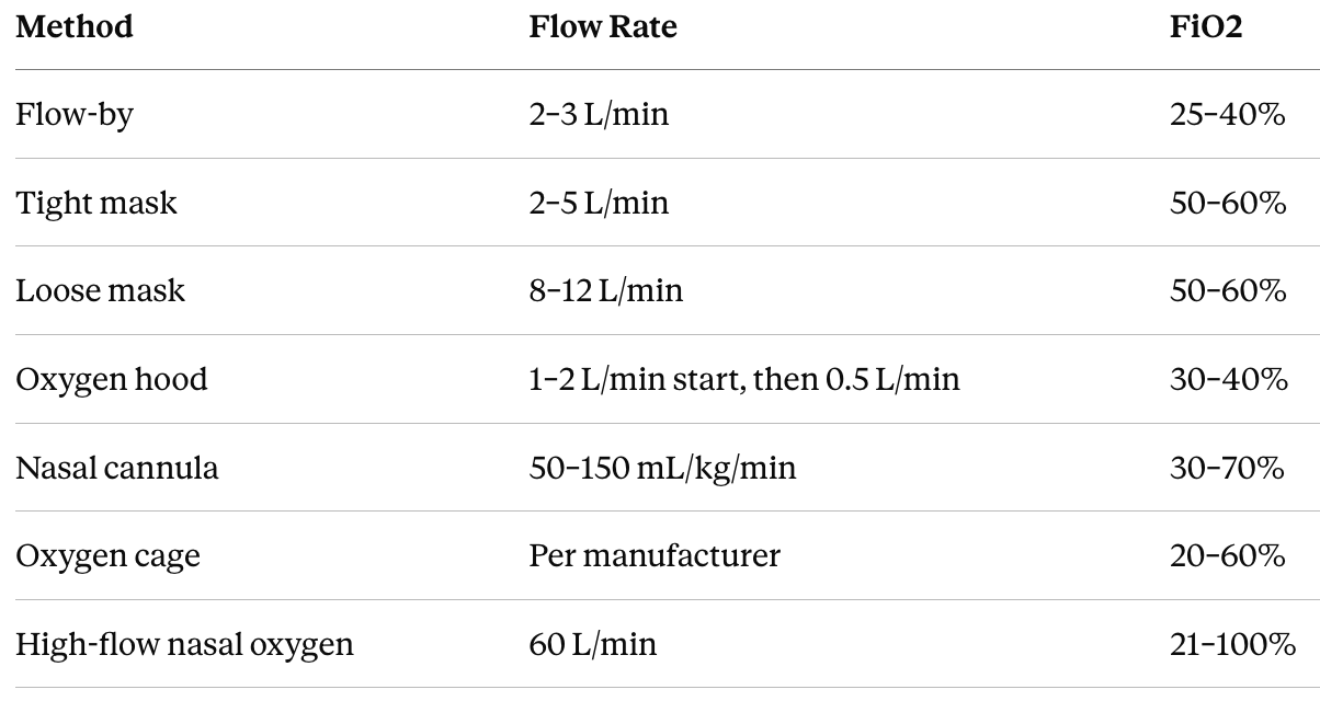
Getting oxygen delivery right — and knowing which monitoring tool to trust — makes the difference in an unstable patient.
A few notes on monitoring: SpO2 is often unreliable, especially in awake, moving, or panting patients, so don't anchor your treatment decisions on a single reading. End-tidal CO2 is most useful once a patient is intubated. Reach for a blood gas specifically when you're monitoring for hypoventilation. And keep in mind that CT requires intubation with breath holds, so tap any pleural effusion or pneumothorax first, before the patient is anesthetized.
Upper Airway Emergencies
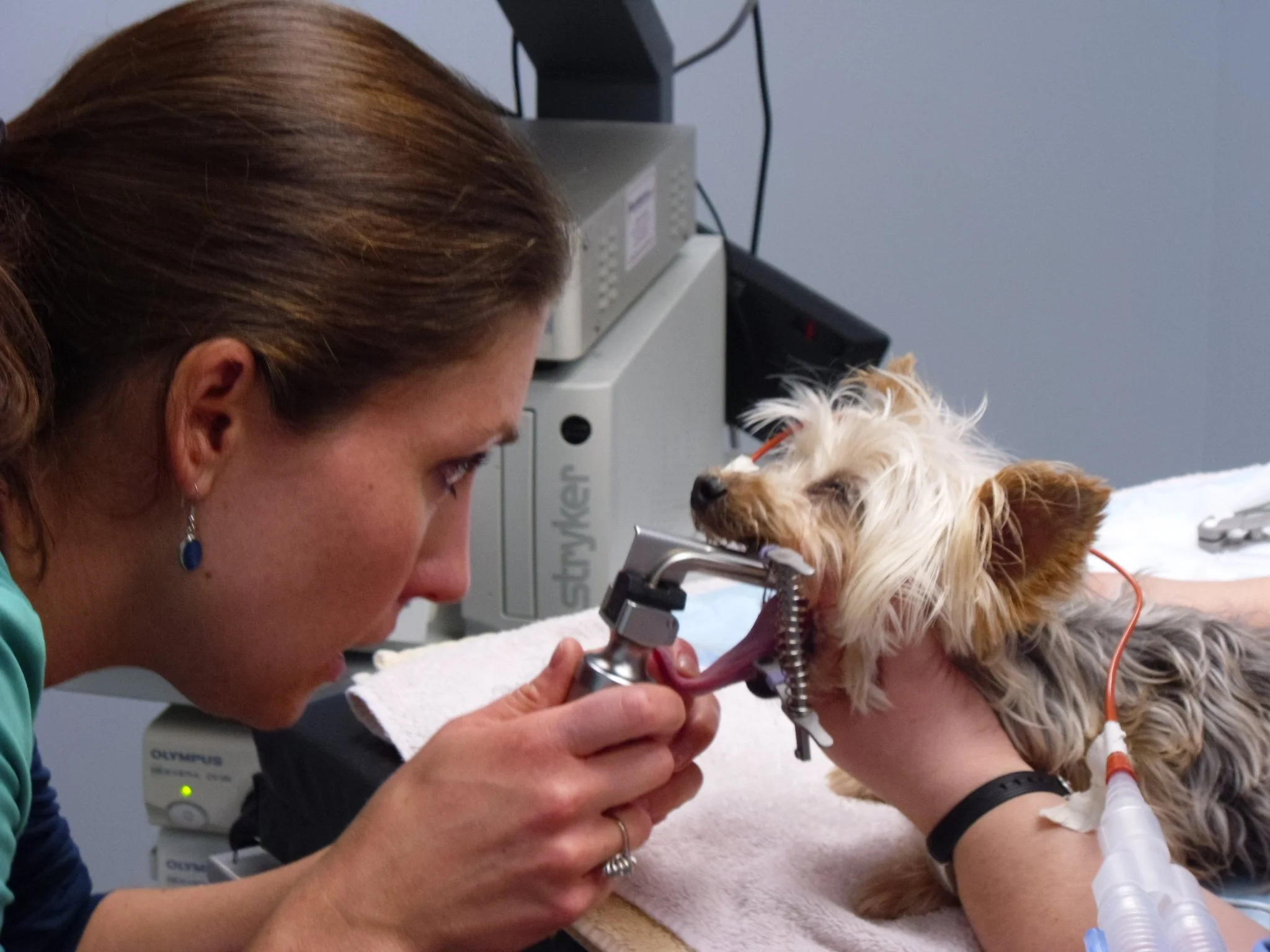
A laryngeal examination after sedation is an important diagnostic step that should not be skipped.
Clinical pattern recognition. Stertor points to nasal pathology; stridor points to upper airway pathology. Static airway sounds suggest a fixed obstruction, while dynamic sounds suggest a functional obstruction — think BOAS, laryngeal paralysis, or tracheal collapse.
Treatment priorities. Start with sedation — torbugesic, acepromazine, dexmedetomidine, and propofol are all reasonable choices depending on the patient in front of you. Steroids are essential for getting airway inflammation under control, and IV fluids plus intubation should follow as needed.
Timing decision. If BOAS surgery is scheduled for the next day, keep the patient intubated and sedated. If surgery will be delayed longer than that, consider a temporary tracheostomy or palatopexy so the patient can be safely extubated in the meantime.
Lung Diseases
Pulmonary edema. Cardiogenic edema — high-pressure, low-protein fluid — calls for oxygen, furosemide, and indicated cardiac medications. Non-cardiogenic edema is increased-permeability edema seen with acute lung injury, contusions, and hemorrhage, and these patients require oxygen and judicious fluids rather than diuretics.
Pulmonary contusions. Oxygen — traditional or high-flow — is the mainstay, with mechanical ventilation reserved for patients who truly need it. Favor small-volume fluid resuscitation with hypertonic saline, adding vasopressors early if needed, and address coagulopathy as needed.
Pneumonia. Pediatric, geriatric, and immunosuppressed patients, along with those who already have upper airway disease, are the ones most at risk. True bacterial pneumonia is genuinely rare in cats; when a cat does develop pneumonia, think fungal, or look for a concurrent airway disease driving it. Treatment starts with oxygen and judicious fluids, adding vasopressors early if the patient is hypotensive.
Don't wait on radiographs to start antibiotics if your clinical suspicion is high enough and favor antibiotics with good lung tissue penetration, like fluoroquinolones, doxycycline, TMS, or chloramphenicol. Ampicillin-sulbactam doesn't technically top that penetration list, but it's still the classic first-line starter unless the patient has been on antibiotics recently — and active pulmonary inflammation can actually improve drug penetration beyond what you'd expect.
On three-view radiographs, look for air bronchograms and sometimes a background interstitial pattern, since patients with pre-existing lung disease like bronchitis or feline asthma are already at higher risk of pneumonia. Add a respiratory PCR panel, especially with relevant exposure history like dog parks or boarding, and fungal testing based on travel or geographic history. A lung wash is reasonable if the patient is stable enough to tolerate it, but warn owners the patient can look worse before the results help you make things better.
Feline Asthma (Feline Lower Airway Disease). These cats show up with increased expiratory effort and an abdominal push, breathing fast and shallow. Some cough or show tracheal sensitivity; plenty don't. Wheezes are often audible on auscultation, and history matters — a smoking household should raise your suspicion. Use TFAST to rule out pleural effusion before you commit to a diagnosis.
Treatment centers on oxygen, butorphanol, and a bronchodilator. Injectable terbutaline is a reasonable option when a mask isn't well tolerated, and a puff of albuterol can double as a diagnostic trial — give it, then reassess respiratory rate about 10 minutes later. Add a short-acting steroid like dexamethasone or dex-SP.
When you genuinely can't tell asthma from CHF and imaging isn't available yet, oxygen, butorphanol, a puff of albuterol, and a single dose of furosemide (2 mg/kg IM) is a reasonable empiric combination — albuterol won't meaningfully harm a CHF cat's heart, and one dose of furosemide won't push a stable cat's kidneys over the edge. Steroids are the exception: a single dose can significantly worsen a cat in CHF, so hold off until you're more confident it isn't cardiac.
On radiographs, look for a diffuse bronchointerstitial pattern with increased bronchial markings — the classic "train tracks and donuts" — along with hyperinflation (lung extending past the last rib) and diaphragmatic flattening. Notably, there's no cardiomegaly or pulmonary venous congestion. Right middle lung lobe consolidation from mucus plugging can occur, and pneumothorax, lung lobe torsion, and bronchiectasis are reported but uncommon.
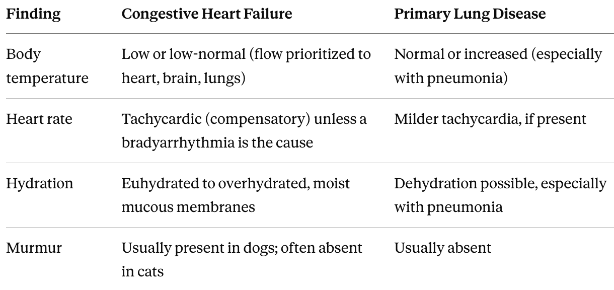
Triage Tips: CHF vs. Primary Lung Disease
When the presentation is ambiguous, these exam and history findings help separate cardiogenic from primary respiratory causes.
Pleural Space Diseases
Work-up. Start with TFAST, fluid PCV/TS, glucose, and lactate, adding PT/PTT and cytology if the effusion is hemorrhagic.
Thoracocentesis technique. Sedation/analgesia and oxygen first. Enter at the 7th to 9th intercostal space, cranial to the rib, to avoid the neurovascular bundle; insert dorsal for air, ventral for fluid. Fenestrating the catheter is not recommended.
Pneumothorax. TFAST is diagnostic with no lung sliding and no B-lines. In the unstable trauma patient, tap before radiographs — don't wait for radiographs to confirm what you can already diagnose at the table. Oxygen itself is genuinely therapeutic here: displacing nitrogen from the alveoli promotes re-expansion.
Diaphragmatic hernia. Positional radiographs, TFAST, or CT can confirm. On exam, watch for an empty abdomen on palpation and borborygmi ausculated in the chest. Support the patient in a Bailey chair or on a ramp, and maintain oxygen support throughout.
Take-Home Pearls
Stabilize before you image: sedation, oxygen, and thoracocentesis (if indicated) come before the radiograph table
TFAST is the fastest triage tool you have — use it early and repeatedly reassess your patient
SpO2 is often unreliable in awake patients; don't rely on it alone
In pulmonary contusions, avoid prophylactic antibiotics and favor small-volume resuscitation over crystalloid boluses
Empirical antibiotics for suspected pneumonia are appropriate before radiographs when suspicion is high
In a cat with respiratory distress and an unclear cardiac-vs-asthma picture, oxygen + butorphanol + albuterol +/- a single dose of furosemide is a reasonable empiric bridge — but hold the steroids until you're more confident it isn't CHF
In the unstable trauma patient with suspected pneumothorax, tap before imaging
Oxygen is directly therapeutic in pneumothorax by promoting alveolar re-expansion
Download the Respiratory Emergencies Top Takeaways (Free)
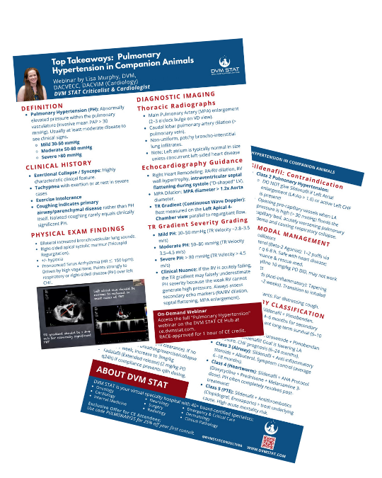
Want a concise, clinic-friendly summary you can save or share with your team?
Download the Respiratory Emergencies Top Takeaways Quick Reference PDF, which includes the oxygen delivery chart, triage comparison table, feline asthma empiric treatment approach, and treatment priorities in an easy, printable format.
Missed the live CE? You can still take the free course on-demand and receive 1 hour of RACE-approved CE here and check out our entire live and on-demand CE library here.
The dyspneic patient rarely gives you time to second-guess the plan. DVM STAT criticalists are available 24/7 for immediate phone consults on unstable patients, and full case reviews are returned in written consult reports within two hours.
Need Help Co-Managing Critical ECC Cases?
Through DVM STAT's Emergency & Critical Care teleconsultations, primary care teams gain access to board-certified criticalists who provide:
Case-specific diagnostic guidance
Step-by-step treatment plans
Support for interpreting complex or conflicting test results
Follow-up support to adjust treatment plans over time with the same criticalist for case continuity
24/7 Consults are available on demand and designed to support the veterinarian-client-patient relationship while improving patient outcomes.
Already have an account? Submit an Emergency & Critical Care teleconsult or call the STAT line directly.