
Pulmonary Hypertension in Companion Animals: Practical Diagnostic and Treatment Guidelines

In DVM STAT’s recent CE webinar on Pulmonary Hypertension, Lisa Murphy, DVM, DACVECC, DACVIM (Cardiology) shared evidence-based guidance on diagnosis, classification, treatment and prognosis of pulmonary hypertension in dogs and cats.
Pulmonary hypertension (PH) is easy to miss in practice. The clinical signs — exercise intolerance, exertional collapse, a soft murmur — overlap with primary respiratory disease and left-sided heart failure. PH has 5 broad classes associated with very different etiologies and prognoses, and getting the classification right changes the treatment plan and the prognosis. In one important case, the classification determines whether a cornerstone of PH treatment is safe to use at all.
Below are top takeaways from the recent CE Webinar on Pulmonary Hypertension with Dr. Murphy.
Missed the live CE? You can still take the full "Pulmonary Hypertension in Companion Animals" course on-demand and receive 1 hour of free RACE-approved CE here and check out our entire live and on-demand CE library here.
What Is Pulmonary Hypertension?
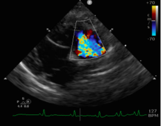
Pulmonary hypertension is abnormally elevated pressure within the pulmonary vasculature — defined as an invasive mean pulmonary arterial pressure (PAP) greater than 30 mmHg. Severity is graded as:
Mild: 30–50 mmHg
Moderate: 50–80 mmHg
Severe: >80 mmHg
Patients usually need at least moderate disease before clinical signs appear. A patient with mild PH on echo may not be the patient with the cough and collapse history in your exam room — so imaging findings need to be interpreted alongside the clinical picture, not in isolation.
Recognizing Pulmonary Hypertension in Practice
Clinical history: The single most characteristic feature is exertional collapse or syncope. Also watch for tachypnea (with exertion, or at rest in severe cases) and exercise intolerance. Coughing is a common owner complaint, but isolated coughing rarely reflects clinically significant PH on its own — it more often points to primary airway or parenchymal disease.
Physical exam findings include:
Bilateral increased bronchovesicular lung sounds
Right-sided apical systolic murmur (tricuspid regurgitation)
+/- hypoxia
Pronounced sinus arrhythmia (HR ≤ 150 bpm), driven by high vagal tone — this finding points strongly toward respiratory or right-sided disease (PH) rather than left-sided CHF
Diagnostic Imaging
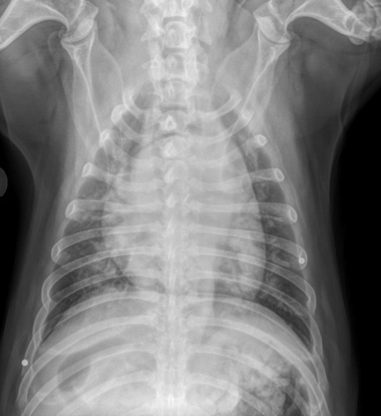
Thoracic radiographs. Look for:
Main pulmonary artery (MPA) enlargement — the classic 2–3 o'clock bulge on the VD view
Caudal lobar pulmonary artery dilation, larger than the accompanying pulmonary vein
Non-uniform, patchy broncho-interstitial infiltrates
Importantly, the left atrium is typically normal in size on radiographs unless there is concurrent left-sided heart disease.
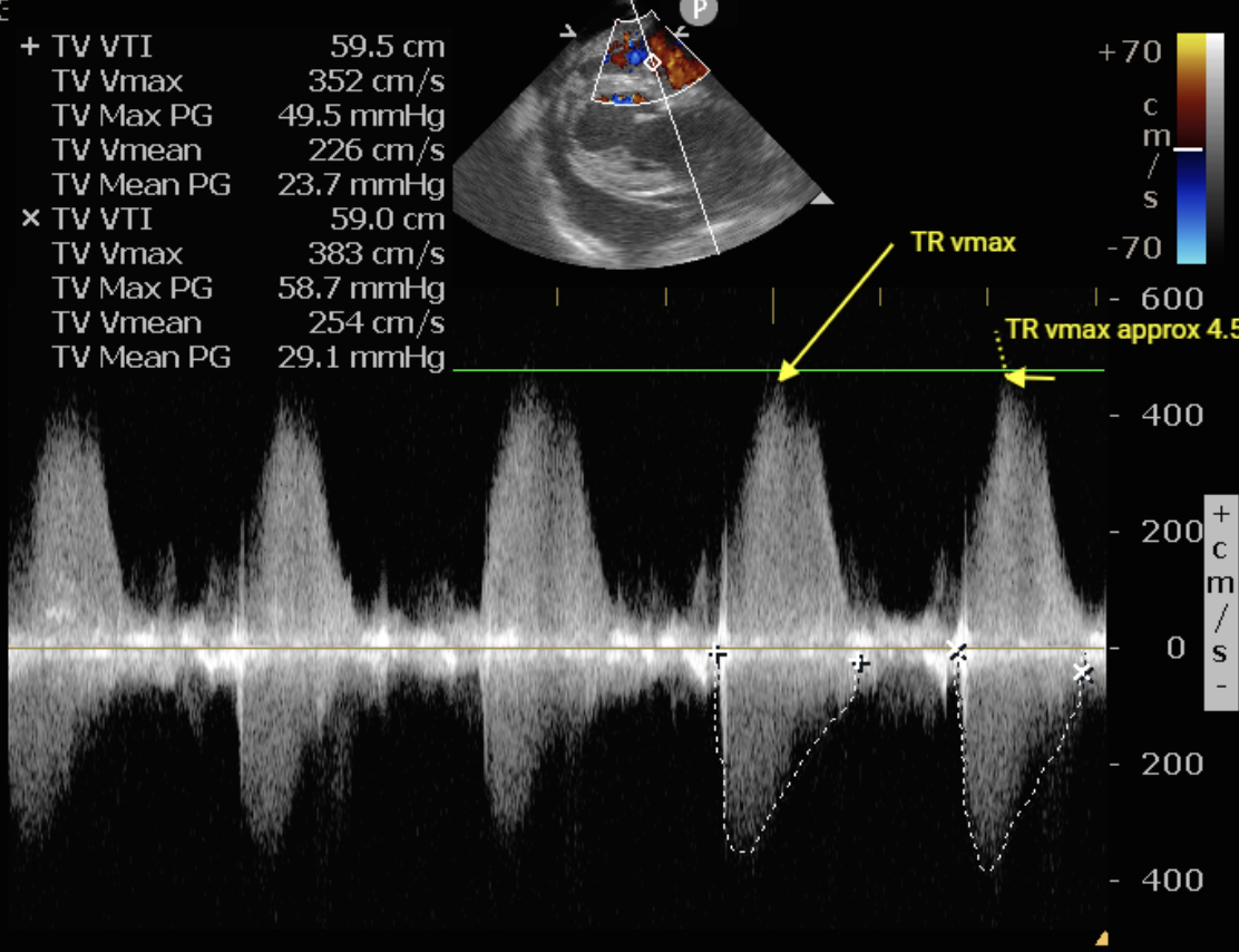
Echocardiography. Echo remains the primary tool for confirming and grading PH.
Right heart remodeling: RA/RV dilation, RV wall hypertrophy, and intraventricular septal flattening during systole (the classic "D-shaped" LV)
MPA dilation: MPA diameter greater than 1.2x the aortic diameter
TR gradient (continuous wave Doppler): best measured from the left apical 4-chamber view, aligned parallel to the regurgitant jet
TR gradient severity grading:
Mild PH: 30–50 mmHg (TR velocity ~2.8–3.5 m/s)
Moderate PH: 50–80 mmHg (TR velocity 3.5–4.5 m/s)
Severe PH: >80 mmHg (TR velocity >4.5 m/s)
if the right ventricle is acutely failing, the TR gradient can falsely underestimate PH severity — a weak RV simply can't generate high pressure. Don't be reassured by a modest TR gradient in a patient who otherwise looks sick. Always cross-check secondary echo markers: RA/RV dilation, septal flattening, and MPA enlargement.
The Five Classifications of Pulmonary Hypertension
Correctly classifying PH is the single most important step in building a safe treatment plan.
Class 1 — Increased flow (shunts): Reverse (right-to-left) shunts such as PDA, VSD, and ASD. This is the #1 cause of PH in cats.
Class 2 — Increased pulmonary venous pressure: Left-sided heart disease with an enlarged left atrium (MMVD, DCM). High LA pressure passively transmits backward into the pulmonary vasculature. Do not treat Class 2 PH with sildenafil (more below).
Class 3 — Increased vascular resistance: Lower airway or parenchymal disease. This is the #1 cause of PH in geriatric dogs.
Class 4 — Infectious / increased vascular resistance: Dirofilariasis (heartworm disease). More common in younger dogs and rescues; causes severe arterial endarteritis and vascular obstruction.
Class 5 — Thromboembolic / increased vascular resistance: Pulmonary thromboembolism (PTE), associated with protein-losing nephropathy/proteinuria, sepsis, neoplasia, and IMHA.
Multimodal Management
Regardless of class, several supportive therapies are commonly layered onto the classification-specific plan:
Bronchodilators. Albuterol (a beta-2 agonist)is safe in patients with concurrent heart disease and useful as both a maintenance and rescue medication. Theophylline is an alternative but may not be effective in every dog.
Corticosteroids. A tapering course of prednisone (1–2 weeks) for anti-inflammatory effect, transitioning to inhaled fluticasone for longer-term control.
Cough suppressants as needed for a distressing cough.
Sildenafil: The Cornerstone Therapy — With One Critical Contraindication
Sildenafil, a PDE-5 inhibitor, produces selective pulmonary vasodilation and reduces RV afterload. It's the backbone of PH treatment for most classes. Sildenafil must be dosed q8h in dogs because of rapid clearance — twice-daily dosing under-treats these patients. If exercise tolerance or breathing has not improved in 7–10 days, dose escalation may be necessary. Tadalafil is a reasonable extended-release alternative when q8h compliance isn't realistic for the household.
The contraindication that matters most: Do not give sildenafil to a patient with Class 2 (left heart disease) PH if there is left atrial enlargement (LA:Ao > 1.6) or active left-sided CHF. Opening the pre-capillary vessels while LA pressure is already high (>30 mmHg) floods the capillary bed, acutely worsening pulmonary edema and precipitating respiratory collapse. This is a case where the intuitive "give the vasodilator" instinct can actively harm the patient, so classification has to come before treatment.
Treatment by Classification
Class 1 (Shunts): Sildenafil + pimobendan. Monitor PCV every 4–6 months for secondary polycythemia. Long-term survival is excellent — typically 5–10 years.
Class 2 (Left heart disease): Furosemide + pimobendan + ACE inhibitor. No sildenafil. The goal is lowering LA pressure. Expected CHF-driven prognosis is 6–24 months.
Class 3 (Airway disease): Sildenafil + anti-inflammatory steroids + albuterol. Focus is symptom control; average survival is 6–18 months.
Class 4 (Heartworm disease): Sildenafil + the AHA adulticide protocol (doxycycline + prednisone + 3-dose melarsomine). PH often resolves completely after treatment.
Class 5 (PTE): Sildenafil + antithrombotics (clopidogrel, enoxaparin) + treatment of the underlying trigger. Acute mortality risk is high, so this class warrants the most aggressive, closely monitored approach.
Take-Home Pearls
Exertional collapse/syncope is the most characteristic clinical sign of PH — isolated coughing is not
A pronounced sinus arrhythmia points toward right-sided/respiratory disease over left-sided CHF
TR gradient can underestimate severity in an acutely failing RV — always assess secondary echo markers
Classification (1–5) determines treatment and prognosis
Never give sildenafil in Class 2 PH with LA enlargement (LA:Ao > 1.6) or active left CHF
Sildenafil must be dosed q8h in dogs due to rapid clearance
Class 4 (heartworm-associated) PH carries the most favorable long-term outlook once the underlying infection is treated
Download the Pulmonary Hypertension Top Takeaways (Free)
Want a concise, clinic-friendly summary you can save or share with your team?
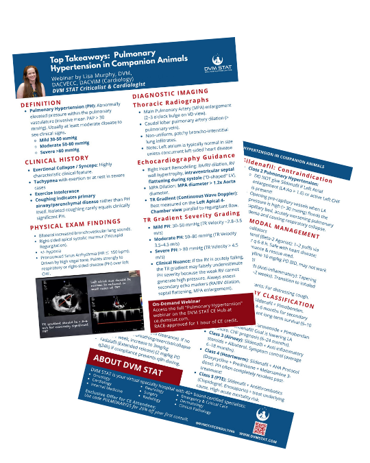
Download the Pulmonary Hypertension Top Takeaways Quick Reference PDF, which includes the classification framework, diagnostic criteria, and treatment protocols in an easy, printable format.
Missed the live CE? You can still take the free course on-demand and receive 1 hour of RACE-approved CE here.
Check out our entire live and on-demand CE library here.
Need Help Co-Managing a Cardiology or Respiratory Case?
Through DVM STAT's Cardiology teleconsultations, primary care teams gain access to board-certified cardiologists who provide:
Cardiology consultations (including radiology, ECG, BP, bloodwork)
Echocardiogram interpretation
ECG assessment
Follow-up support to adjust treatment plans over time with the same specialist for case continuity
Consults are available on demand and designed to support the veterinarian-client-patient relationship while improving patient outcomes.
Already have an account? Submit a Cardiology teleconsult or call the STAT line directly.